
Figure 1. Cerebral blood flow (CBF) changes and acute injury during middle cerebral artery occlusion (MCAO) in young and aged SHR.
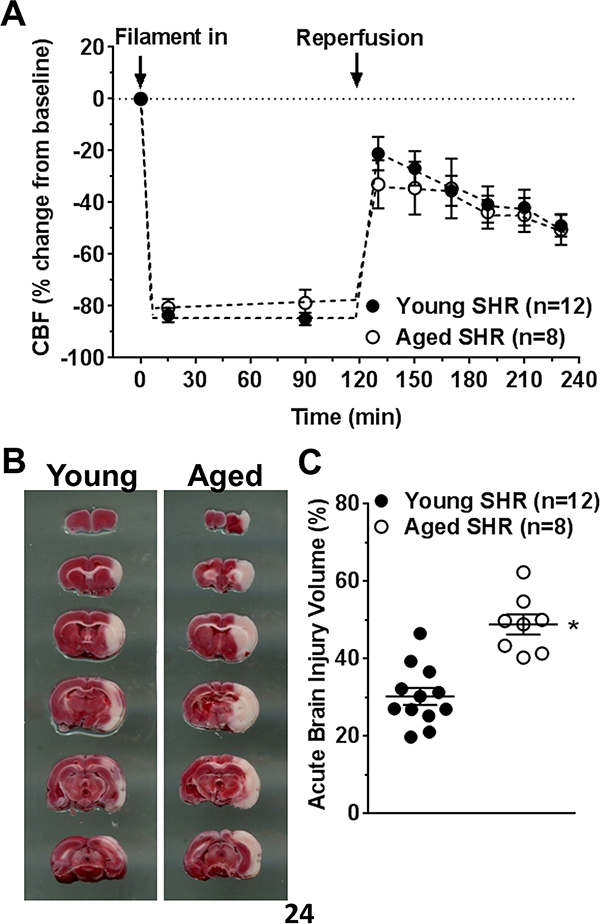
(A) Percent change in CBF during MCAO and reperfusion. Both young and aged SHR had similarly decreased CBF during filament occlusion and similar reperfusion that was incomplete in both groups. (B) Representative coronal sections stained with TTC and used for injury volume analysis. (C) Graph showing acute injury volume was increased in aged SHR. * p<0.05 vs. young SHR by Student’s t-test.