
Figure 3. Erythrocyte aggregation and hemoglobin staining within the brain injury area and the role of PAI-1.
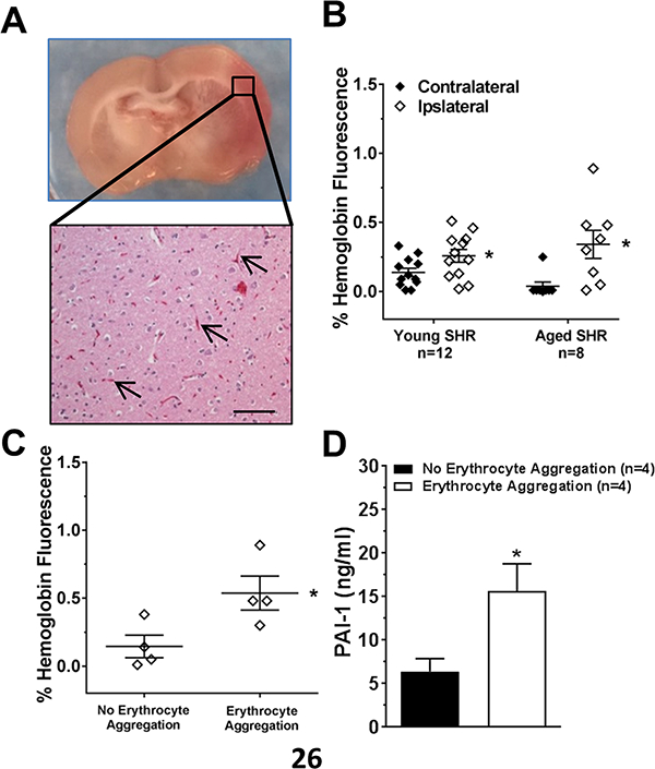
Representative images of a brain coronal section with pink coloration within the injury area (A, upper) and erythrocyte aggregation in brain capillaries with H&E staining (A, lower) in aged SHR. Scale bar: 100 μm (B) Graph showing quantification of hemoglobin staining by immunohistochemistry within the contralateral and ipsilateral side of occlusion for young and aged SHR. Hemoglobin was increased in both groups on the ipsilateral side. * p<0.05 vs. contralateral by Student’s t-test. (C) Graph showing comparison between aged SHR brains with and without pink coloration (erythrocyte aggregation). Brains with pink coloration had increased hemoglobin. * p<0.05 vs. no erythrocyte aggregation by Student’s t-test. (D) Comparison of PAI-1 in serum of aged SHR with and without pink coloration. * p<0.05 vs. no erythrocyte aggregation by Student’s t-test.