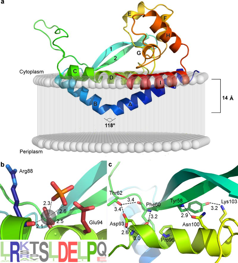
FIGURE 1. PglC reveals a distinct architecture and topology for monotopic membrane proteins.
a, Predicted position of PglC with respect to the membrane, including the reentrant membrane helix (RMH) formed by the helix-break-helix motif of helices A and B (N to C termini colored blue to red). b, Depiction of the PglC active site showing the conserved Asp–Glu dyad with Mg2+ and phosphate ligands and sequence logo. c, The AHABh (alpha-helix-associated beta-hairpin)-motif that defines the superfamily fold is formed by a β-hairpin comprising β-strands 1 and 2 packing against helix D.

