
Figure 6. Gain of function of SIRT3 in OA chondrocytes eliminated pre-existing mtDNA4977 deletion mutation and improved mitochondrial function.
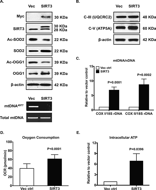
Cultured primary human knee OA chondrocytes were transfected with pCDNA4-Myc-HisB-Sirt3 and vector control for 48 hours. Expression of SIRT3 was confirmed by Western blot with antibodies to either Myc or SIRT3 (A, top panel). Acetylation and expression of SOD2 and OGG1 (A, top panel), and expression of subunits of mitochondrial respiratory complex III and V (B) were analyzed by Western blot. The mtDNA4977 deletion mutation (A, bottom panel), mtDNA content (C), oxygen consumption (D) and intracellular ATP (E) were determined as described in Figure 2 legend. Data in A and B represent 3 individual experiments with 3 different OA donors (2 females and 1 male, age 81, 58 and 64, respectively). Data in C, D and E were the mean of 4 individual experiments with 4 different OA donors (1 male and 3 females, age 67, 70, 56 and 68, respectively). Student t-test was used for statistical data analysis in C, D and E (n=3, 3–4 replicates for each donor) comparing vector control with SIRT3 overexpression. The mean differences, 95% CIs and p values for COXI/18S rDNA and COXII/18S rDNA: 6.106 ± 0.5714, 4.833 to 7.379, p<0.0001, and 7.747 ±0.7820, 6.004±9.489, p<0.0001, respectively. The mean differences, 95% CIs and p values for oxygen consumption and ATP levels: 55 ± 2.678, 11.16 to 21.95, p<0.0001, and 5.874 ± 1.205, 2.529 to 9.219, p=0.0082, respectively.