
Figure 1. Platelets express the anaphylatoxin receptor C3aR.
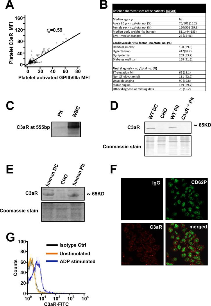
(A) Analyzing patients with coronary artery disease, expression of C3aR and activated GPIIb/IIIa (activation specific Ab PAC-1) was measured by flow cytometry. The correlation of C3aR with activated GPIIb/IIIa is depicted. Spearman`s rank coefficient rs=0.59, p<0.001, n=501 patients. (B) Baseline characteristics of the analyzed patients (C) Expression of C3aR on resting human platelets and WBC as control was detected by isolation of mRNA and RT-PCR of transcribed cDNA with specific primers. A C3aR specific band could be detected at 555bp on a 2% agarose gel. (D+E) Protein expression of C3aR on thrombin- stimulated (0.01 U/ml) murine (D) and human (E) platelets was analyzed by western blot. CHO cells served as negative control, murine bone marrow derived dendritic cells (DC) or human monocyte derived dendritic cells served as positive control. One of 3 representative blots is depicted. Coomassie Blue staining of the PVDF membranes after blotting served as loading control. (F) ADP-stimulated (20 µM) human platelets in PRP were fixed with 1% PFA and spun on poly-L-lysine coverslips. Permeabilized platelets were analyzed for expression of CD62-P (green) and C3aR (red) with a Zeiss LSM 800 microscope (63x objective. Area of interest cropped 4.7x) (G) Human platelets were isolated, stimulated with ADP (20 µM) and analyzed by flow cytometry for surface expression of C3aR. n=8, p<0.05 vs. resting platelets, one representative diagram is shown.