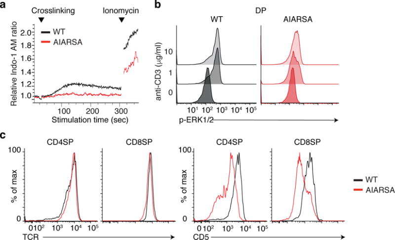
Fig. 4. Mutation of the PIPRSP motif in LAT impedes TCR signal transduction of DP thymocytes.

(a) Flow cytometric analysis of calcium-sensitive fluorescence changes of CD45.2+mCherry+ DP T cells, loaded with Indo-I AM and stimulated with crosslinked anti-CD3 over time. (b) Flow cytometric analysis of phosphorylated Erk in CD45.2+mCherry+ DP thymocytes, in response to crosslinked anti-CD3 stimulation. (c) Flow cytometric analysis of surface expression of the TCR (left) or CD5 (right) in CD45.2+ mCherry+ CD4SP or CD8SP thymocytes. Experiments were repeated independently twice with four independent animals and yielded similar results.
