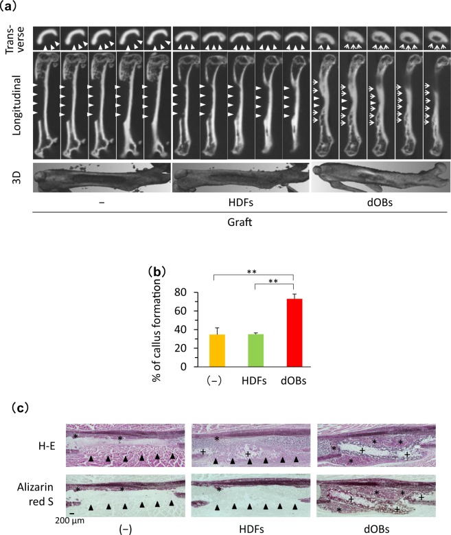
Figure 4.
Bone healing was facilitated by transplantation of the fibronectin-coated NanoCliP-FD gel with dOBs. Fibronectin-coated NanoCliP-FD gel with HDFs or dOBs was prepared as in Fig. 3, and transplanted into an artificial segmental bone defect lesion that was created at femoral diaphysis in NOG/SCID mice. Control mice were not transplanted (−). Mice were sacrificed 21 days after the surgery. (a,b) µCT images of the femur were acquired. Serial 10-µm slices (top and middle) and 3D reconstructed (bottom) images (a) and %Callus formation (b) are shown. (c) Serial sections of the tissues were stained with H-E (upper) and Alizarin red S (lower). In (a) triangles and arrows represent bone defect lesions and regenerated bone tissue, respectively. In (b), values are means ± SD. N = 3 mice. **p < 0.01 vs. non-transplantation control. In (c), *and +represent regenerated bone tissue and NanoCliP-FD gel, respectively, and arrowheads represent bone defect lesions.

