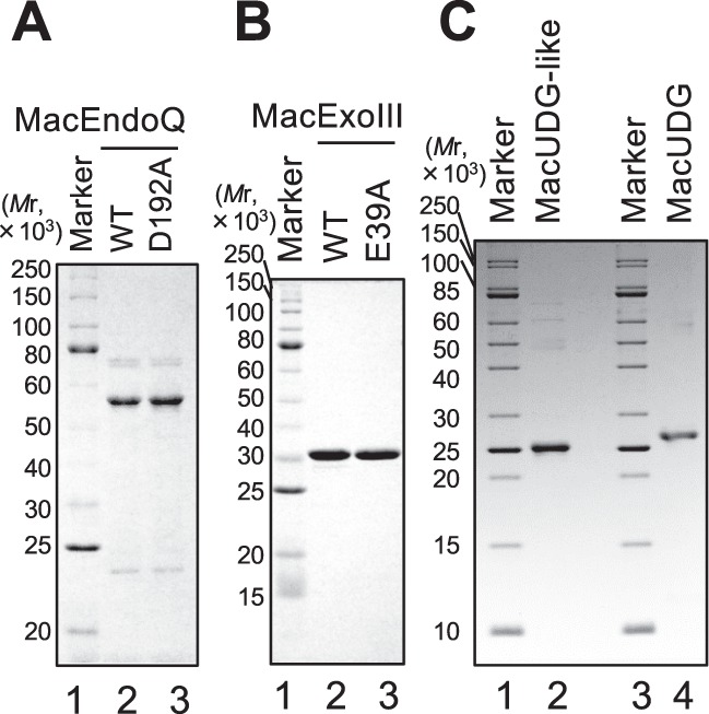
Figure 1.

Preparation of recombinant proteins. Each purified protein (1 μg) was run on 12% (A,B) or 15% (C) SDS-PAGE, followed by CBB staining. The molecular weights of the markers are shown on the left of the panels. (A) Lane 1, protein marker (NEB, P7703); lane 2, MacEndoQWT (MW: 54111.1); lane 3, MacEndoQD192A (MW: 54067.1). (B) Lane 1, protein marker (NEB, P7703); lane 2, MacExoIIIWT (MW: 32548.5); lane 3, MacExoIIIE39A (MW: 32490.4). (C) Lanes 1 and 3, protein marker (NEB, P7704); lane 2, MacUDG-like (MW: 25231.11); lane 4, MacUDG (MW: 25298).
