
Figure 5.
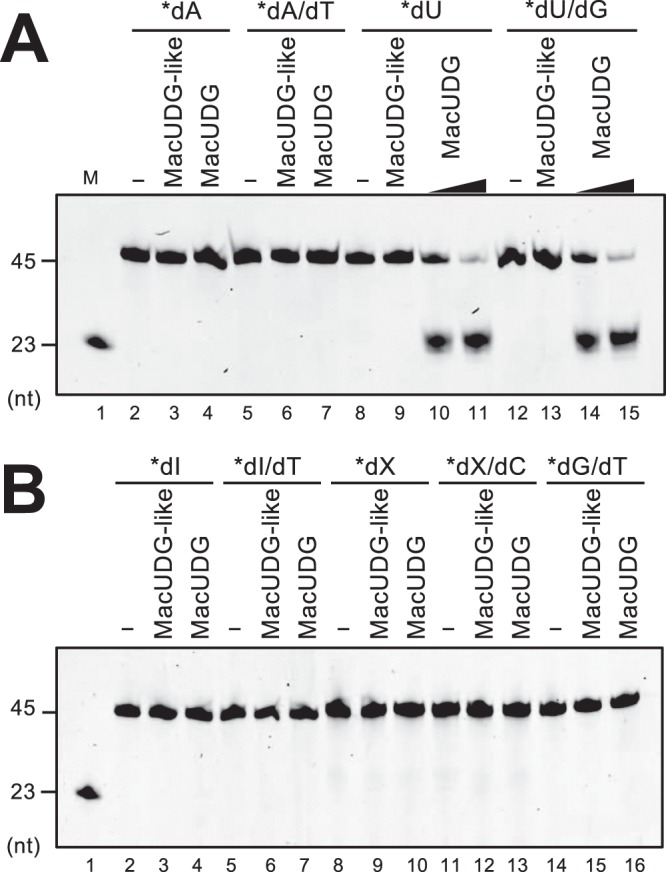
MacUDG exhibits glycosylase activity specific for uracil while MacUDG-like is inactive. 5′-Cy5-labeled ssDNA (A: lanes 2–4, 8–11; B: lanes 2–4, 8–10) or dsDNA (A: lanes 5–7, 12–15; B: lanes 5–7, 11–16) were incubated without (A: lanes 2, 5, 8, 12; B lanes 2, 5, 8, 11, 14) or with 1 μM MacUDG-like (A: lanes 3, 6, 9, 13; B: lanes 3, 6, 9, 12, 15), 1 nM MacUDG (A): lanes 10, 14), and 1 μM MacUDG (A: lanes 4, 7, 11, 15; B: lanes 4, 7, 10, 13,16) at 37 °C for 10 min. The products were treated with alkaline and heating, which cleaved DNA at AP site. DNA substrates are indicated at the top of the panels; dA, normal ssDNA; dA/dT, normal dsDNA; dU, dI, and dX, damaged ssDNA; dU/G, dI/T, and dX/C, damaged dsDNA; dG/dT, mismatched dsDNA. Asterisks represent Cy5-labeled strands. Cleavage products were separated by 8 M urea-10% PAGE. M, DNA marker (A,B, lane 1). The cropped gels are used in the figure, and the full-length gels are presented in Supplementary Fig. S11.