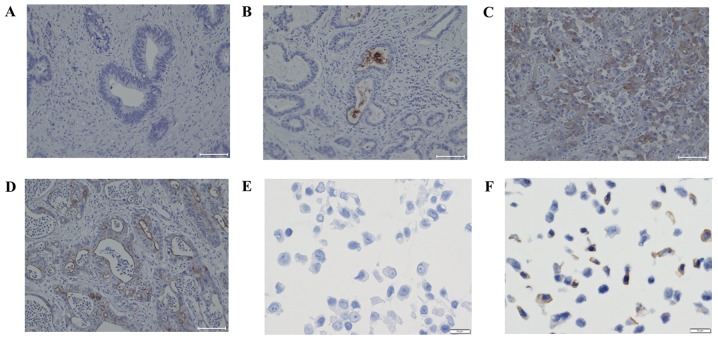
Figure 1.
Representative immunohistochemical staining of CD133 in EHBDCA and GBCA cells. (A) No stain for CD133 in tumor cells. (B) Luminal surface and intraluminal cell stain for CD133 in tumor cells. (C) Cytoplasmic stain for CD133 in tumor cells. (D) Mixed stain for CD133 in tumor cells. Scale bar, 100 µm. (E) MCF-7 cells were used as a negative control and (F) MDA-MB-468 cells were used as a positive control. Scale bar, 20 µm. CD133, prominin-1; EHBDCA, extrahepatic bile duct cancer; GBCA, gallbladder cancer.

