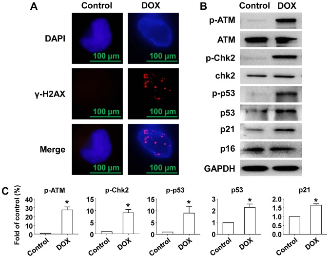
Figure 2.
Effects of DOX on γ-H2AX foci formation and expression of DNA damage related proteins in RPMI-8226 cells. RPMI-8226 cells were incubated with/without 100 nM DOX for 24 h. (A) Cells were fixed and stained with γ-H2AX (red) and DAPI (blue) at the indicated time points. (B) RPMI-8226 cells treated with/without 100 nM DOX for 24 h were extracted and proteins were incubated with primary antibodies against p-ATM, ATM, p-Chk2, Chk2, p-p53, p53, p21 and p16. GAPDH was used as a loading control. (C) Protein quantification was conducted and data were shown with fold of control. Data were obtained from at least three independent experiments. The significance of differences between two groups was determined by Student's t-test. *P<0.05 vs. control. DOX, doxorubicin; MTT, multiple myeloma; ATM, ataxia telangiectasia mutated.

