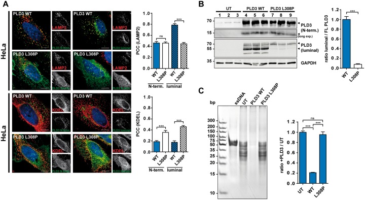
Figure 1.
Impaired lysosomal delivery, proteolytic processing and loss of function of the PLD3-L308P mutation. (A) Confocal microscopy of HeLa cells transfected with the PLD3-WT or PLD3-L308P mutation followed by staining with the indicated antibodies. Pearson correlation coefficient (PCC) for co-localization for the indicated antibodies is depicted; n = 12 cells. (B) Immunoblot of PLD3-WT or PLD3-L308P mutation with indicated PLD3-specific antibodies and GAPDH. Quantification of the fragments relative to full-length protein is shown. UT = untransfected. (C) Ethidium-bromide stained polyacrylamide gel analysis of untreated ssDNA substrate and treated with untransfected HeLa cell lysates or transfected with PLD3-WT or PLD3-L308P-mutation transfected HeLa cell lysates. Quantification represents n = 3. ***P < 0.001; ns = not significant.

