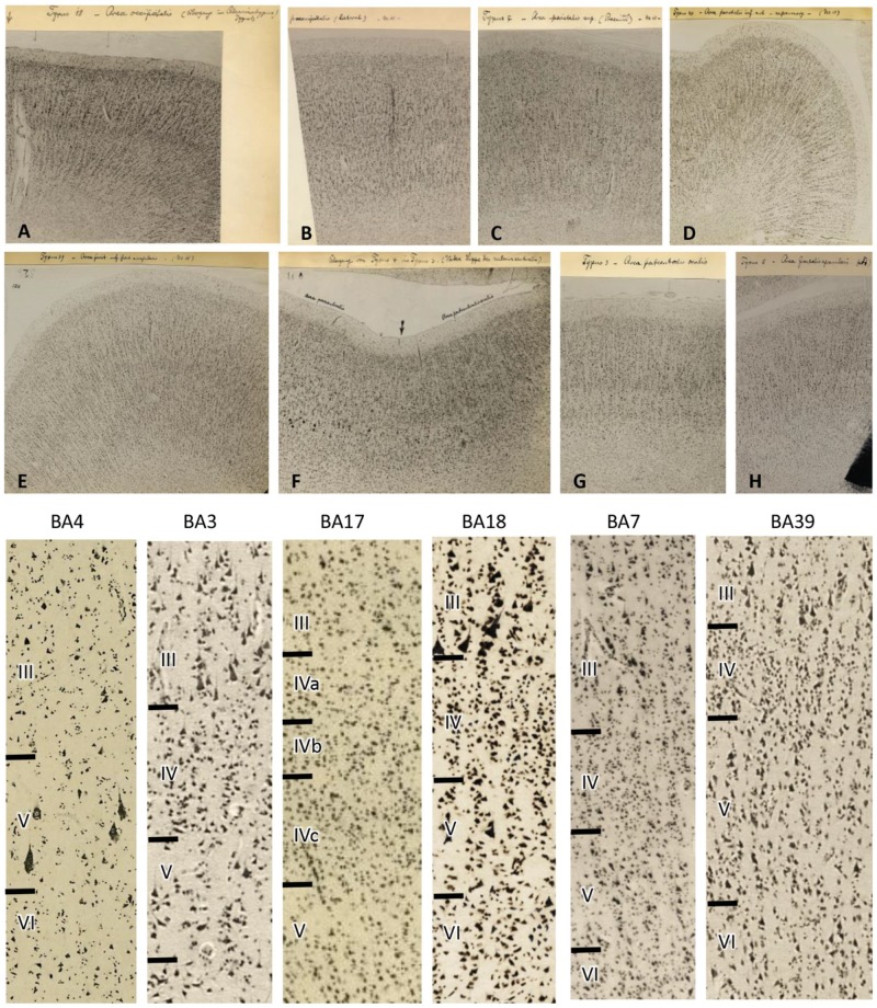
Figure 6.
Original micrographs of human cortical areas by K. Brodmann (specimen M15). Transcriptions of Brodmann’s hand writing: (A) Area occipitalis BA18, secondary visual cortex at the transition to BA17. (B) Praeoccipitalis BA19 (higher visual cortex). (C) Area parietalis sup. (precuneus ant) BA7. (D) Area parietalis inf. ant = supramarg BA40, inferior parietal cortex. (E) Area pariet. post. inf = angularis BA39, temporo-occipital cortex. (F) Transition from BA4 to BA3, (primary motor to primary somatosensory cortex). (G) Area postcentralis oralis BA3, primary somatosensory cortex. (H) Area frontalis agranularis BA6, premotor cortex. Bottom: Higher magnifications of the original micrographs (A–H) demonstrate the larger (or at least equally large) pyramidal cells in layer III compared with those cells in layer V. This cytoarchitectonic feature is called ‘externopyramidization’ and typical for higher unimodal (BA18 as an example in the visual system) and multimodal (examples BA7 and BA39) areas. In contrast, the primary motor (BA4), somatosensory (BA3), and visual (BA17) cortices display larger pyramidal cells in layer V than III. Note the wider layer IV in primary sensory (BA3 and BA17) than multimodal (BA7 and BA39) areas, and the lack of a clearly visible layer IV in the primary motor area (BA4). With permission of the C. & O. Vogt Archive, Institute of Brain Research, University Düsseldorf.

