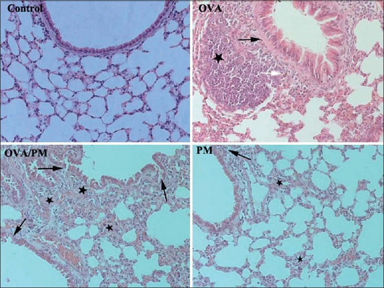
Figure 5.

Comparison of the changes in lung tissues of mice in different groups (hematoxylin and eosin, ×200). The control group had intact terminal bronchioles and alveolar epithelia and showed no inflammation. The OVA group showed hyperplasia of smooth muscles of the small bronchi (black arrow), hyperplasia of lymphatic follicles (star), and infiltration of eosinophils (white arrow). The OVA/PM group displayed mild loss of tracheal epithelial cells, widening of the alveolar septa (black arrows), infiltration of inflammatory cells, and hyperplasia of smooth muscles of the small bronchi (stars). The PM group displayed the presence of inflammatory cells in peribronchiolar regions (black arrow) and substantial alveolar epithelial hyperplasia (stars). PM: Particulate matter; OVA: Ovalbumin.
