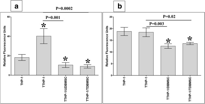
Fig. 8.
The adhesion of monocytes (THP-1) to HUVEC was evaluated by measuring THP-1 fluorescence intensity using a fluorescence microplate reader. THP-1 were initially cultured alone (THP-1) or with 100 μM H2O2 (TTHP-1) for 24 h and then cultured with DBMSCs (5:1 THP-1:DBMSC ratio) that were initially cultured alone (UDBMSC) or with 100 μM H2O2 (TDBMSC) for 24 h. After 24 h culture, THP-1 were labelled with 5 μM green fluorescent cell tracker stain CMFDA and added to HUVEC monolayer (HUVEC were initially cultured with or without 100 μM H2O2 for 24 h). As compared to untreated THP-1, the adhesion of TTHP-1 to H2O2 untreated HUVEC significantly increased while the adhesion of TTHP-1/UDBMSCs and TTHP-1/TDBMSC to H2O2 untreated HUVEC significantly reduced after 30 min (a). As compared to TTHP-1, the adhesion of TTHP-1/UDBMSCs and TTHP-1/TDBMSC to H2O2 untreated HUVEC significantly reduced after 30 min (a). As compared to untreated THP-1, the adhesion of TTHP-1 to H2O2 pretreated HUVEC was not significantly changed (P > 0.05) after 30 min while the adhesion of TTHP-1/UDBMSCs and TTHP-1/TDBMSC to H2O2 pretreated HUVEC significantly reduced after 30 min as compared to untreated THP-1 and TTHP-1 (b). Each experiment was performed in triplicate and repeated for five times with five independent preparations of DBMSCs and HUVEC. *P < 0.05. Bars represent standard errors

