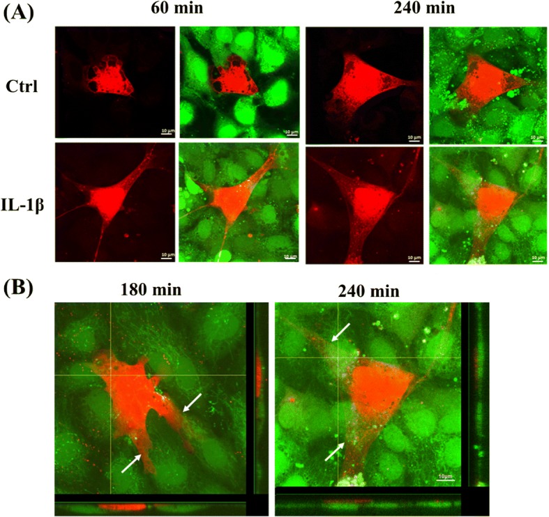
Fig. 3.

Effects of IL-1β in morphology and interaction between HUVECs and MSCs. Labeled MSCs with CellTracker™ Orange were seeded on HUVECs stained with Calcein AM and co-cultivated for 30 to 240 min. After a period of 60 min, MSCs attached to HUVECs and the morphology were still spherical but developed form of cytoplasmic offshoot. a After 60 min, MSC became flattened and adhered to HUVEC monolayer. IL-1β promoted adhesion (left, 60 min) and transendothelial migration abilities (right, 240 min) of MSCs. b After 180 and 240 min, MSCs extended long plasmic filopodia and integrated into the HUVEC monolayer. Orthogonal projections illustrate that MSCs inserted into HUVEC monolayer (left, 180 min) and formation of filopodia caused transendothelial migration (right, 240 min). Arrows indicate MSC migration through the HUVEC. Horizontal bar: XZ plane of confocal image stack; vertical bar: YZ plane of confocal image stack. Scale bar = 10 μm
