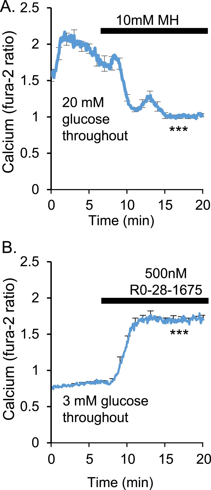
Figure 1.
[Ca2+]i levels depend on glucokinase activity. (A) In high glucose (20 mM), acute exposure with a 10-mM concentration of the glucokinase inhibitor MH caused a rapid decrease in [Ca2+]i to levels typically observed in very low glucose (n = 17 islets, ***P < 0.001). (B) Stimulation of glucokinase activity with 500 nM Ro-28-1675 in low glucose (3 mM) led to constitutively high [Ca2+]i (n = 10 islets, ***P < 0.001).

