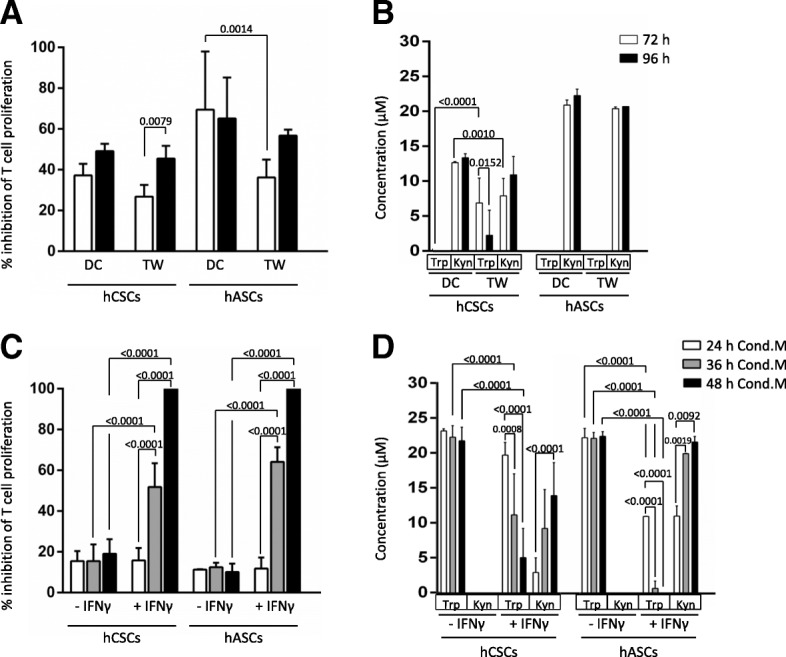
Fig. 4.

Human cardiac/stem progenitor cells (hCSCs) inhibit T lymphocyte proliferation via a paracrine mechanism. a CFSE-labeled hPBMCs were stimulated with PHA and cultured alone, in direct contact (DC), or in a transwell setting (TW) with hCSCs (ratio 1:10 hCSCs:hPBMCs). b Concentrations of tryptophan (Trp) and kynurenine (Kyn) were determined by HPLC in the supernatants. c CFSE-labeled hPBMCs were stimulated with PHA and cultured alone or in conditioned medium (Cond.M.) from hCSCs cultures activated or not with interferon (IFN)-γ. Conditioned media were generated for 24 h (white bars), 36 h (grey bars), and 48h (black bars). d Concentrations of Trp and Kyn were determined by HPLC in the conditioned media. Proliferation of the viable population of CD3 T lymphocytes (CD3+/7AAD–) was assayed by loss of CFSE staining after 72 h (white bars) and 96 h (black bars) for TW and DC experiments (a) and after 96 h for Cond.M. experiments (c). Percentage of cells per generation and percentage of inhibition of proliferation was determined using FSC Express software against proliferation of activated hPBMCs alone. Human adipose-derived mesenchymal stem cells (hASCs) were used as a positive control for T cell proliferation inhibition (ratio 1:25 hASCs:hPBMCs). Adjusted p values are shown
