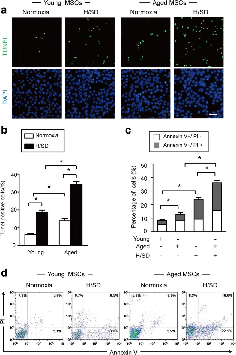
Fig. 2.

Hypoxia significantly increased apoptosis in aged MSCs. a Representative immunofluorescence images of terminal deoxynucleotidyl transferase-mediated nick-end labeling (TUNEL) (green fluorescence) and 4,6-diamidino-2-ph under normal conditions and after hypoxia/serum deprivation (H/SD). b Quantification of the rate of apoptosis of BM-MSCs is shown as the percentage of apoptotic cells. c Quantification of apoptosis is shown as the percentage of cells (with marker of annexin in early and late apoptotic stages). Early apoptosis: annexin V+/PI−; late apoptosis: V+/PI+. Data are expressed as the means ± SEM; n = 5; *p < 0.05. d Representative results of the FACS analysis in BM-MSCs under normal conditions and H/SD viable cells: annexin V−/PI−; early apoptosis: annexin V+/PI−; late apoptosis: V+/PI+; necrotic: V−/PI+
