
Figure 1.
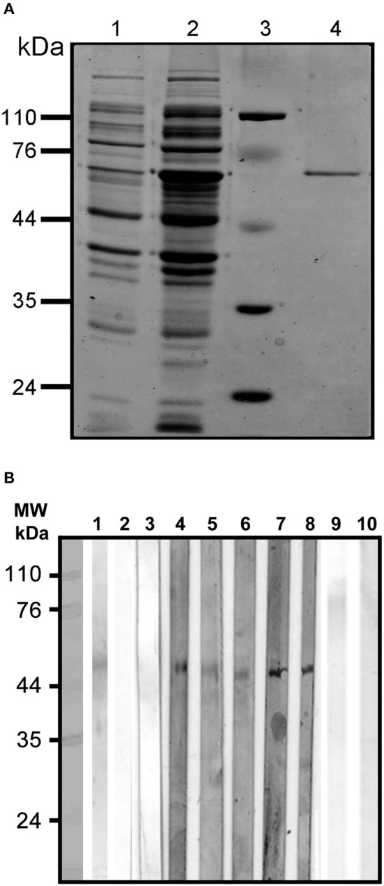
(A) Purification under denaturing conditions of rEhCRT. Proteins were visualized by 12% SDS-PAGE and Coomassie staining; (1) cell lysate with plasmid, (2) cell lysate with plasmid after IPTG induction, (3) molecular weight, (4) rEhCRT eluid from Ni-NTA Agarose column, molecular weight marker are indicated in kilodaltons. (B) Westernblot of rEhCR. The reactivity of the rEhCRT was tested against sera from the different groups studied; (1) 5 μg of rEhCRT electrotransfered to NC membrane, (2–3) serum from control individuals (NEG), (4–6) serum of AP-ALA patients, (7–8) R-ALA patients, and (9–10) reactivity against E. coli-LPS antibody.