Figure 4.

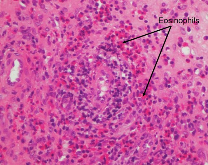
Sinus biopsy demonstrating vessels with lymphocytic infiltrates and marked accumulation of eosinophils in extravascular areas (cells with intensely pink cytoplasm, arrow).

Sinus biopsy demonstrating vessels with lymphocytic infiltrates and marked accumulation of eosinophils in extravascular areas (cells with intensely pink cytoplasm, arrow).