Fig. 1.
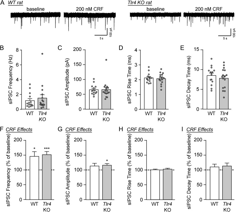
CRF increased GABA transmission in the CeA of WT and Tlr4 KO rats. (A) Representative sIPSC traces from CeA neurons of WT and Tlr4 KO rats at baseline and during CRF (200 nM) superfusion. (B–E) Baseline sIPSC frequencies (B), amplitudes (C), rise times (D) and decay times (E) were similar across both genotypes (WT: 14 cells from three rats; Tlr4 KO: 16 cells from five rats). (F) CRF significantly increased the sIPSC frequencies in WT rats (12 cells from three rats; one-sample t-test, t(11) = 2.86, *P < 0.05) and Tlr4 KO rats (14 cells from five rats; t(13) = 4.84, ***P < 0.001). There was no difference in the magnitude of drug effects between groups (unpaired t-test, t(24) = 0.31, P = 0.76). (G) The mean sIPSC amplitude was increased by CRF in Tlr4 KO rats (one-sample t-test, t(13) = 3.01, *P < 0.05), though the drug’s effects were not significantly different across genotypes (unpaired t-test, t(24) = 0.27, P = 0.79). (H–I) CRF had no effect on sIPSC kinetics across both genotypes. All data are presented as mean ± SEM.

