Figure 3.
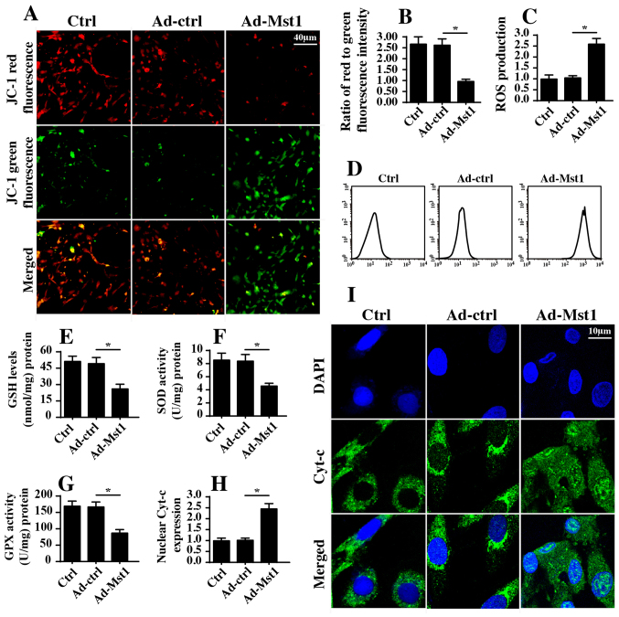
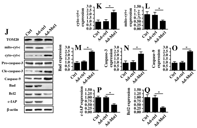
Mst1 overexpression is associated with mitochondrial damage. (A and B) Mitochondrial potential was observed via JC-1 staining. Red fluorescence indicated normal mitochondrial potential, whereas green fluorescence indicated damage to mitochondrial potential. (C and D) ROS production was measured by flow cytometry. Ad-Mst1 transduction increased the ROS content in A549 cells. (E-G) Antioxidants were detected by ELISA. (H and I). Cyt-c translocation into the nucleus was observed by co-staining cells with cyt-c and DAPI. (J-Q) Western blotting of proteins associated with mitochondrial apoptosis. The expression levels of pro-apoptotic and anti-apoptotic proteins were analyzed by western blotting. TOM20 was used as a loading control for mito cyt-c expression. *P<0.05. Ad, adenovirus; Bad, Bcl2-associated death promoter; Bcl2, B-cell lymphoma 2; c-IAP, cellular inhibitor of apoptosis protein; cyt-c, cytochrome c; GPX, GSH peroxidase; GSH, glutathione; mito, mitochondrial; Mst1, mammalian STE20-like kinase 1; ROS, reactive oxygen species; SOD, superoxide dismutase; TOM20, translocase of the outer membrane 20.


