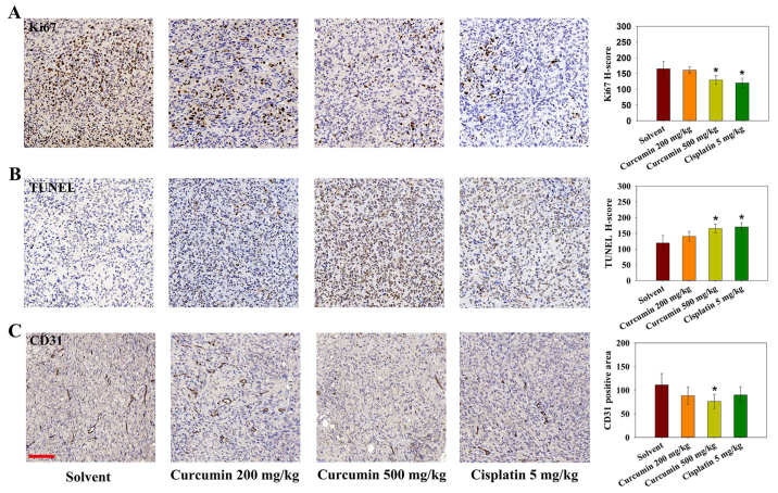
Figure 6.
Effects of curcumin on angiogenesis, cell proliferation and apoptosis in tumor-bearing mice, as determined by Ki67, TUNEL and CD31 staining. Representative histological micrographs of (A) Ki67, (B) TUNEL and (C) CD31 staining. (A and B) Ki67 and TUNEL staining was semi-quantitatively evaluated by H-score. (C) CD31 was semi-quantified by determining the positive area percentage. Data are expressed as the means ± standard deviation. There were 4-5 mice in each experimental group. Scale bar, 100 µm. *P<0.05 vs. the solvent group. CD31, cluster of differentiation 31.

