Figure 1.
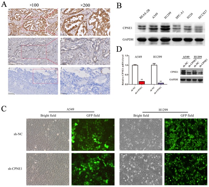
CPNE1 is frequently overexpressed in lung adenocarcinoma tissues and cell lines. (A) A total of 128 formalin-fixed and paraffin-embedded NSCLC tissues were subjected to immunohistochemical analyses of the CPNE1 protein. Representative images are presented of CPNE1 antibody staining in (a and b) NSCLC tissues (c) and normal tissues, at ×100 magnification, and at ×200 magnification (d-f). (B) The level of CPNE1 in human NSCLC cells was detected by western blotting. (C) Lentiviral transfection efficiency was detected using fluorescence microscopy at ×100 magnification to observe cells expressing green fluorescent protein at 5 days following transfection. (D) CPNE1 mRNA and protein levels in stable A549 and H1299 cells. Each cell line was divided into the following two groups: The sh-NC group (cells infected with Lv-si-CTRL) and the sh-CPNE1 group (cells infected with Lv-si-CPNE1). **P<0.01 vs. sh-NC. CPNE1, Copine 1; NSCLC, non-small cell lung cancer; sh-NC, negative control cells; sh-CPNE1, CPNE1-silenced cells.

