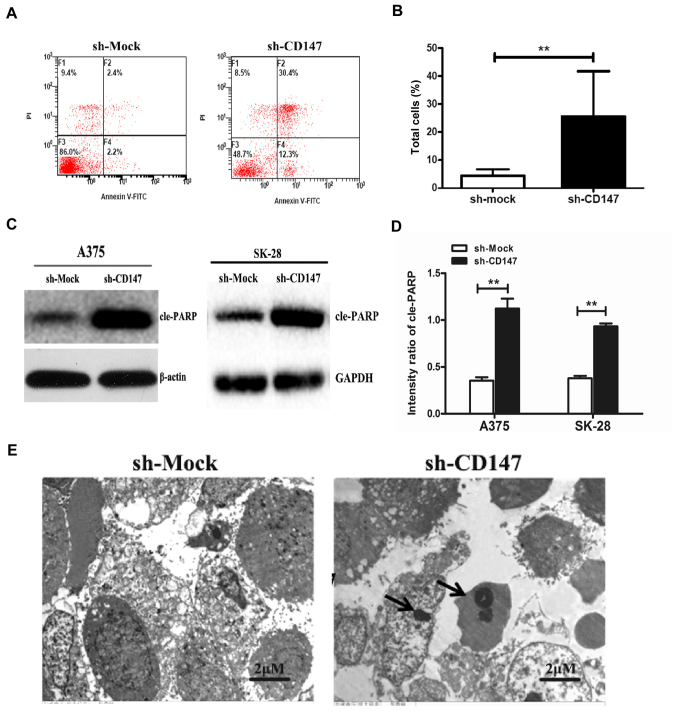
Figure 2.
Inhibition of CD147 promotes the apoptosis of melanoma cell lines. (A) Representative dot plots of Annexin V-FITC and PI staining. After A375 cells were stably transfected with sh-Mock (left) or sh-CD147 vector (right), apoptosis analysis was performed by flow cytometry. (B) Apoptotic rate of sh-CD147- and sh-Mock-transfected A375 cells. The percentage of apoptotic cells was significantly higher with decreased CD147 expression. (C and D) Protein expression levels of apoptosis-associated cle-PARP in sh-CD147- and sh-Mock-transfected A375 and SK-28 cells. (E) Transmission electron microscopy images of apoptosis of sh-CD147-transfected and sh-Mock-transfected A375 cells. Apoptotic bodies are indicated by arrows. n=3 for each experiment. Scale bar=2 μm. **P<0.01. CD147, cluster of differentiation 147; cle-PARP, cleaved poly (ADP-ribose) polymerase; FITC, fluorescein isothiocyanate; PI, propidium iodide; sh, short hairpin RNA.

