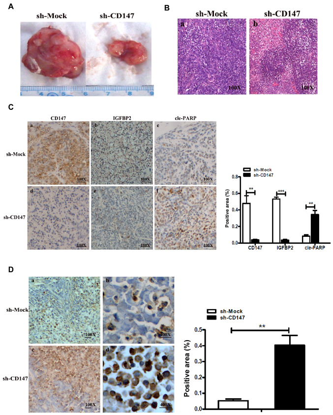
Figure 5.
CD147 with IGFBP2 mediates the apoptosis of melanoma cells in vivo. (A) Tumor volume in the xenograft mouse model generated by inoculating melanoma sh-CD147 A375 and sh-Mock A375 cells. **P<0.05 or ***P<0.01 vs. sh-Mock. (B) Pathological characteristics of xenograft mouse tumors were observed by HE staining. Magnification, ×100. (C) Representative immunohistochemical staining for CD147, IGFBP2 or cle-PARP in serial sections of sh-CD147-injected xenograft mouse tumors or sh-Mock tumors. Magnification, ×100. The area positive for CD147, IGFBP2 and cleaved PARP- in sh-CD147 or sh-Mock cell-injected xenograft mouse tumors was determined by using Image-Pro Plus 6.0 software. (D) Representative TUNEL assay in sh-CD147-injected xenograft mouse tumors and sh-Mock tumors; the apoptosis-positive regions are indicated in brown-yellow. The histogram indicated the TUNEL-positive area (%) (right). Magnifications, ×100 or ×400. CD147, cluster of differentiation 147; cle-PARP, cleaved poly (ADP-ribose) polymerase; IGFBP2, insulin-like growth factor-binding protein 2; sh, short hairpin RNA; TUNEL, terminal deoxynucleotidyl-transferase-mediated dUTP nick end labeling.

