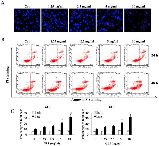
Figure 2.
GLP induces apoptosis of PC-3 cells. (A) Cells were treated with various concentrations of GLP for 24 h. Hoechst 33342 staining was used to analyze apoptotic cells. Magnification, 200×. (B) GLP (0-10 mg/ml) induced apoptosis of PC-3 cells at 24 and 48 h. Apoptosis was quantified by Annexin V/fluorescein isothiocyanate flow cytometry. (C) Percentage of early and late apoptotic cells, as induced by GLP in PC-3 cells. Data are presented as the means ± standard error of the mean. *P<0.05; **P<0.01 compared with the control group (one-way analysis of variance with Dunnett's correction). GLP, Ganoderma lucidum polysaccharides; PI, propidium iodide.

