Figure 1.
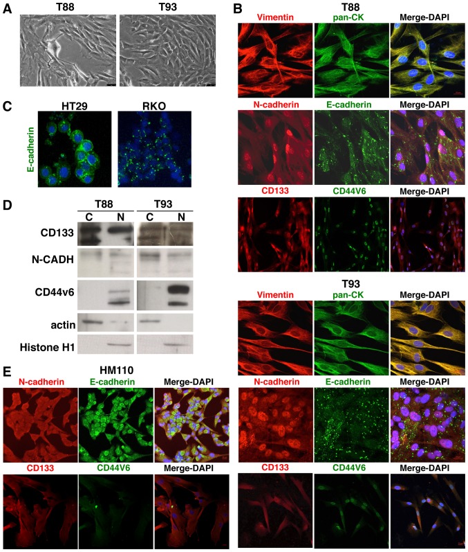
Nuclear N-cadherin, CD133 and CD44v6 localisation in T88 and T93 mesenchymal colorectal cancer (CRC) cells. (A) Brightfield images of T88 and T93 cells at ×10 magnification. (B) Confocal images of T88 and T93 primary colon cancer cells stained for anti-Vimentin (red)/anti-pan-CK (green), anti-N-cadherin (red)/anti-E-cadherin (green) and anti-CD133 (red)/anti-CD44v6 (green) antibodies. Nuclei were counterstained with DAPI (blue). (C) Confocal images of HT29 and RKO cells stained with anti-E-cadherin (green) antibody. Nuclei were counterstained with DAPI (blue). (D) Western blot analysis of cytoplasmic 'C'' and nuclear 'N' extracts from T88 and T93 cells using anti-CD133, anti-N-cadherin, anti-Cd44v6, anti-actin and anti-histone H1 antibodies. Histone H1 and actin were used as nuclear and cytoplasmic marker proteins, respectively. Each lane was loaded with 50 μg of cytoplasmic or nuclear extract. The cropped blots are used in the figure. (E) Confocal images of primary cells from the healthy mucosae of a patient affected by sporadic CRC (HM110). Cells were stained with anti-N-cadherin (red)/anti-E-cadherin (green) and anti-CD133 (red)/CD44v6 (green). Nuclei were counterstained with DAPI (blue).

