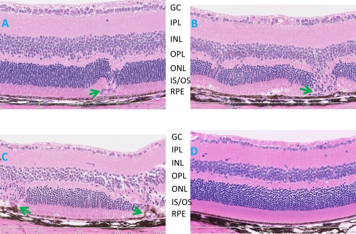
Figure 3.
Histological evaluation of B6.Cg-Crb1rd8Jak3m1J/Boc mice (A–C) demonstrating disease progression at (A) 3 weeks, (B) 2 months, (C) 8 months, and (D) an 8-month-old C57BL/6J control. Aberrant vascular lesions (arrows) are observed as early as 3 weeks, the earliest time examined, and a thinning of the ONL, especially in those areas where the ectopic blood vessels were found. GC, ganglion cell layer; IPL, inner plexiform layer; IS/OS, inner segment/outer segment; ONL, outer nuclear layer; OPL, outer plexiform layer; RPE, retinal pigment epithelium.

