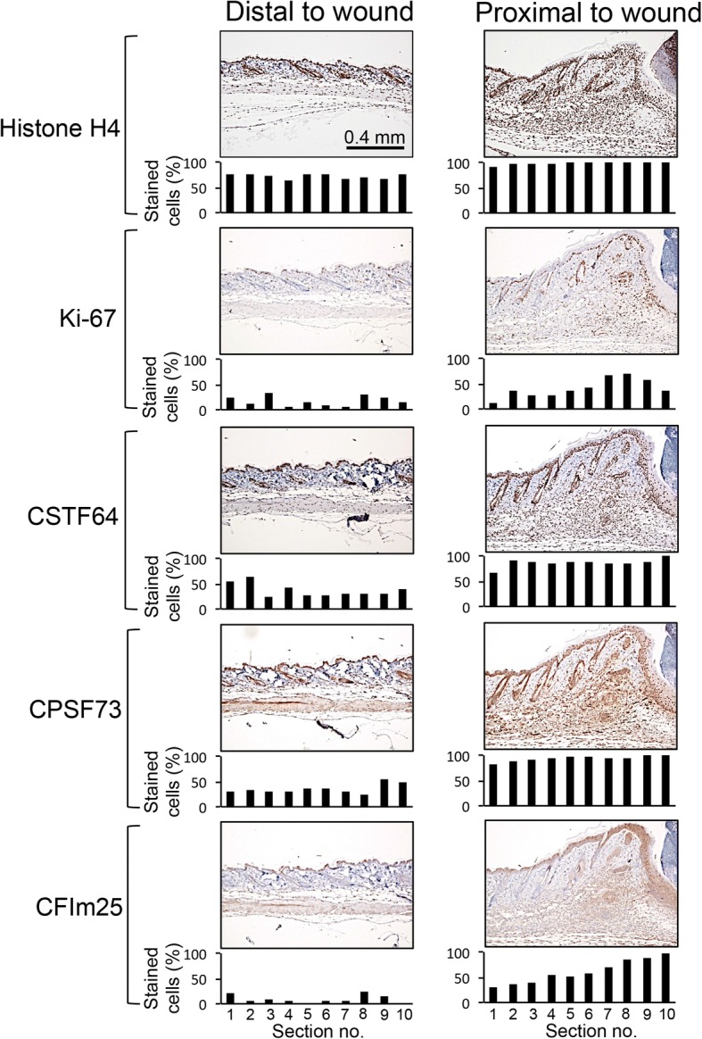
Fig. 7.

Cleavage and polyadenylation factors are expressed at higher levels in fibroblasts near a wound than in fibroblasts of healthy skin. Mouse skin was collected 5 days after introduction of a punch biopsy. Normal mouse skin was collected 2 cm away from the wound. Samples were stained with immunohistochemistry for proliferation marker Ki-67, histone H4 as a control, or alternative polyadenylation and cleavage factors CstF-64, CPSF73 or CFIm25 (brown). Samples analyzed with immunohistochemistry were counterstained with hematoxylin (blue nuclei). Individual cells at different positions from the wounds were assigned positive or negative staining and the percentages are shown. Ki-67 does not label all dividing cells, and likely underestimates the fraction of cells that are actively cycling [122]. Levels of all three cleavage and polyadenylation factors were higher in the fibroblasts, myofibroblasts and immune cells proximal to a wound than in the fibroblast-rich dermal areas of healthy skin distal to the wound
