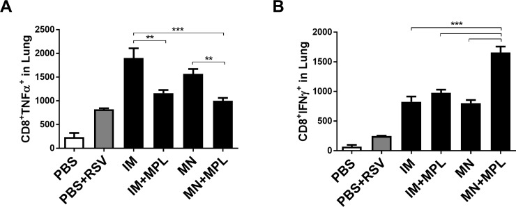
Fig 9. Lung CD8 T cells secreting TNF-α or IFN-γ cytokines as determined by intracellular cytokine staining.
Intracellular cytokine staining of lung cells was carried out by Flow cytometry after in vitro stimulation with F85–93 peptides, a known CD8 T cell epitope, and followed by staining with CD45 and CD8 surface marker antibodies and intracellularly with cytokine antibodies (A) TNF-α. (B) IFN-γ. The Y axis indicate CD8+cytokine+ average cell numbers per lung per mouse in the groups. Mouse groups are the same as described in Fig 3. The data are representative of duplicate experiments. Results are presented as mean ± SEM. Statistical significances were performed by one-way ANOVA and Tukey’s multiple-comparison tests in GraphPad Prism; *** p<0.001, **p<0.01.

