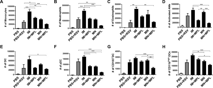
Fig 11. Cellular infiltrates into the lungs are low or prevented by MN patch delivery of FI-RSV after RSV challenge.
Cells from lungs collected at 5 days after challenge were stained with cell type-specific marker antibodies and analyzed by flow cytometry. Results (n = 5) are presented as mean ± SEM and representative of duplicate experiments. The Y axis indicate average infiltrating each designated phenotypic cell numbers in the lung tissue per mouse in the groups. Mouse groups are the same as described in Fig 3. Statistical significances; *** p<0.001, **p<0.01, *p<0.05.

