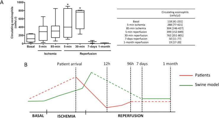
Fig 2. Dynamics of circulating eosinophils in swine subjected to reperfused myocardial infarction (MI).
Whole blood was isolated from the experimental model at basal and at different time points of the ischemia and reperfusion process: after 5-min and 85-min of ischemia (immediately before reperfusion) as well as 5-minutes, 30-minutes, 7-days, and 1-month after reperfusion post-MI. Samples were incubated with FITC-CD45 and PE-CD16 and afterwards measured using flow cytometry. (A) Eosinophil counts in whole blood were analysed. Data were expressed as median with the interquartile range (n≥5 independent experiments) and were analysed by Kruskal-Wallis analysis followed by Dunn’s test. *P<0.05 vs. basal. (B) Dynamics of peripheral eosinophils in ST-elevation MI-patients (red) and in the swine model (green). The drop in peripheral eosinophils detected in ST-elevation MI-patients 12h after coronary reperfusion might be preceded by their progressive increase during ischemia and soon after reperfusion, as observed in the experimental model.

