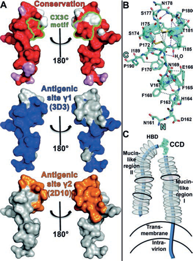
Fig. 4. Sequence conservation within antigenic sites γ1 and γ2, atomic interactions within the RSV G CCD, and model of RSV G glycoprotein.

(A) Top: Surface representation of RSV G with amino acids colored according to conservation. Atoms from the main-chain and conserved side chains are red, similar side chains are pink, and non-conserved side chains are white. The five CX3C motif amino acids are outlined in green. Middle and bottom: the epitope footprints of antigenic site γ1 (bnmAb 3D3 epitope) and antigenic site γ2 (bnmAb 2D10 epitope), respectively. (B) Structure of RSV G161−197 with hydrogen bonds shown in purple dashes and representative sidechain-sidechain hydrophobic interactions (≤4.0 Å) shown in gray dashes. (C) Schematic of membrane-bound RSV G. N- and O-linked glycans are shown by black and gray discs, respectively.
