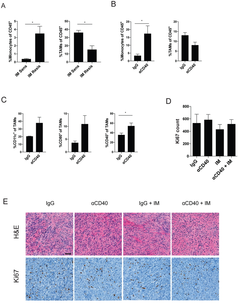
Figure 4. KIT inhibition may be needed for tumor response.
(A) Monocytes and TAMs, as a percentage of CD45+ cells, in the imatinib (IM) sensitive (KitV558Δ/+) and imatinib resistant (KitV558Δ;T669I/+) tumors. KitV558Δ;T669I/+ tumors were analyzed by flow cytometry 3 days after anti-CD40 (αCD40) and assessed for (B) intratumoral monocytes and TAMs, as a percentage of CD45+ cells, and (C) TAMs were assayed for expression of CD11c, CD80, and CD40 expression. KitV558Δ;T669I/+ mice were treated with a single αCD40 injection on day 0, followed by continuous imatinib on day 3. (D) Ki67 count representing the number of positively stained nuclei in one 800 × 730 μm field per tumor. (E) Representative H&E and Ki67 IHC. Bar represents 50 μm. Data represent 2 experiments, mean ± SEM, *P < 0.05 using Student’s t test.

