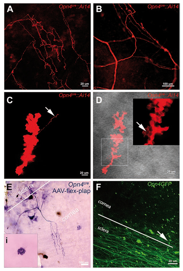
Fig. 1.

Melanopsin expression in cornea revealed by genetically driven markers. (A–D) Corneal epithelium from adult Opn4cre::Ai14 mice. Red color shows melanopsin-driven tdTomato reporter. (A) Confocal imaging in stromal layer of the cornea reveals a network of tdTomato-positive fibers. (B) Small area of tdTomato filled fibers across cornea of another Opn4cre::Ai14 mouse. This figure is an expanded view of the square outlined in Sup. Fig. 1. Image captured between the epithelial and stromal layers reveals bulbous “clump” of cells. Arrow points to a fiber emanating from the clump. (D) tdTomato staining reveals bulbous terminal ending in more superficial layer of cornea than shown in (C). Arrow in the insert denotes localization of tdTomato in the intra-epithelial spaces. Gray background shows bright field view of the corneal epithelium. (E) The cornea from an Opn4cre mouse that received an intravitreal injection of AAV-flex-plap shows the expression of PLAP in the nerve fibers and dendritic-like cells at the level of the corneal endothelium (inset). (F) GFP-positive fibers in sclera and cornea in an Opn4GFP mouse. Scale bars are marked. Comparable findings for all genetic approaches mentioned above were found in 4 to 10 mice across at least 3 litters.
