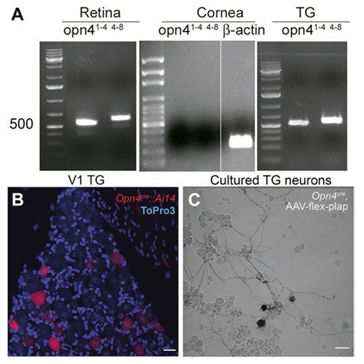
Fig. 3.

Melanopsin mRNA is detected in retina and trigeminal ganglia but not in cornea. Melanopsin-driven markers can be observed in cells imaged in whole-mounted trigeminal ganglia and in culture. (A) RT-PCR results illustrates the expression of the melanopsin mRNA in the retina and the trigeminal ganglion (TG) but not the cornea. The melanopsin expression was probed with two sets of primers encompassing exons 1 through 4 (superscript 1–4) and exons 4 through 8 (superscript 4–8). (B) V1 part of the trigeminal ganglion from Opn4cre::Ai14 mouse (P98). Red color shows Ai14 reporter; blue color shows nuclei of cells (ToPro3 stain). Scale bar: 20 μm. (C) Neuronal culture from the trigeminal ganglion of the Opn4cre mouse infected with the AAV-flex-plap. Black color shows the expression of PLAP. Scale bar: 50 μm. Each PCR result was obtained in no fewer than three mice.
