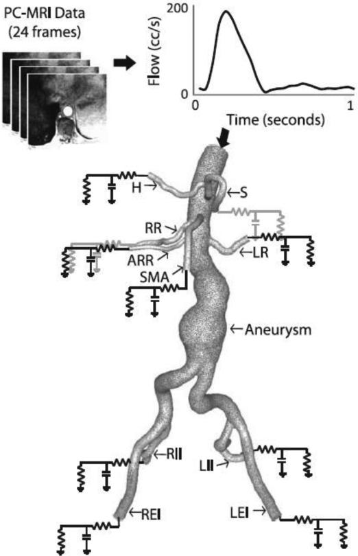
Figure 3.
A patient-specific SC flow waveform measured by phase-contrast MRI (resting conditions) is mapped to the inflow face using a Womersley velocity profile. A patient-specific 3-element Windkessel model with a proximal resistance (Rp), capacitance (C), distal resistance (Rd) boundary condition is used to represent the resistance and compliance of the vasculature downstream of each outlet. The outlet labels are as follows: S=splenic artery, LR=left renal artery, LEI=left external iliac artery, LII=left internal iliac artery, RII=right internal iliac artery, REI=right external iliac artery, SMA=superior mesenteric artery, ARR=accessory right renal artery (Note: accessory renals are not found in the majority of patients), RR=right renal artery, H=hepatic artery. Please note that typically (as in this patient), the splenic and hepatic arteries branch from a common celiac trunk.

