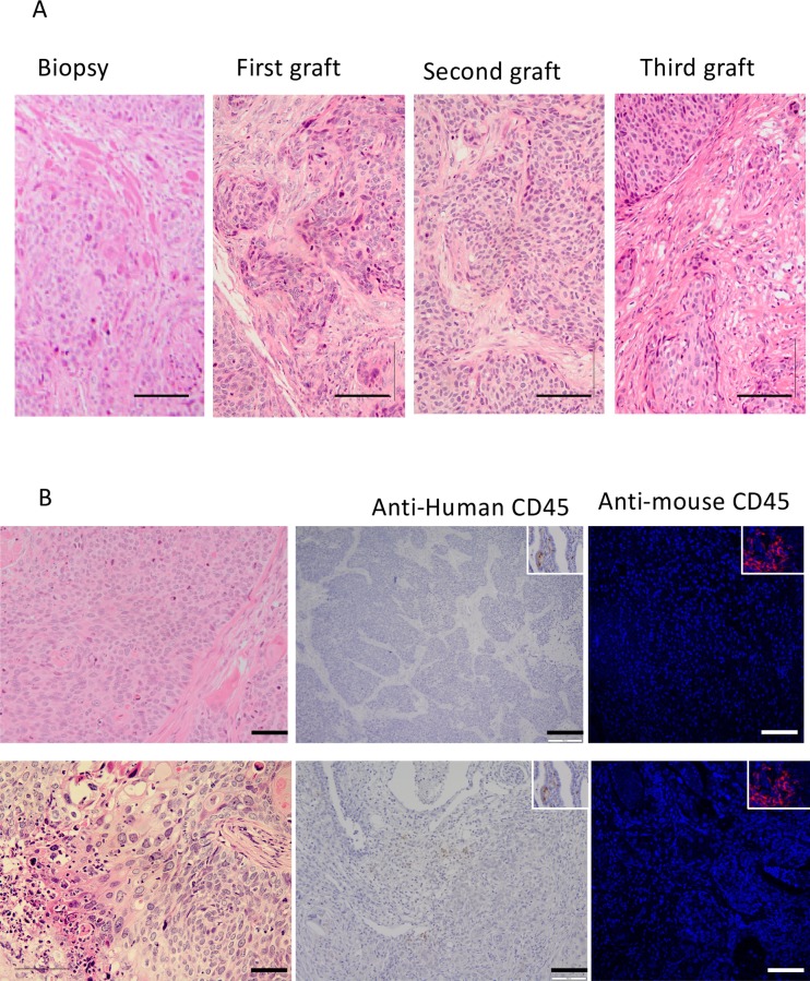
Fig 2. Morphology of serially transplanted cervical cancer PDXs.
A) representative example of an H&E stained cervical squamous cell carcinoma sample showing morphology of the tumour biopsy, primary, secondary and tertiary PDXs. B) Typical examples of negative staining for anti-human CD45 staining (second column), and anti-mouse CD45 (third column). Insets show examples of CD45 positive staining in human cervix biopsies and mouse kidney Scale bars 50 μm.

