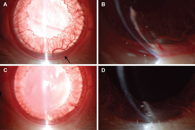
Figure 1. LAU-0901 prevents hypopyon formation in the anterior segment.

Slit lamp photography of the anterior segment revealed hypopyon formation (arrow), increased prominence of iris vessels, and reduced dilation in animals that received LPS treatment (A). Magnified image reveals the hypopyon in the anterior segment, as well as a similar aggregate in the ciliary sulcus behind the iris (B). However, when LPS treatment was accompanied by LAU-0901 administration, no hypopyon was observed and there was mimimal engorgement of iris vessels (C and D).
