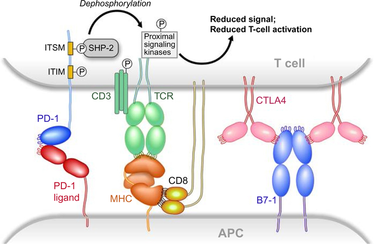
Fig. 16.
Structures of the B7/CD28 family. Structures are modeled on the crystal determinations. Loops have been added to one end of the IgV domains to emphasize the orientation of the CDR-like loops and their interaction with ligand or lack thereof. Reproduced with permission from Freeman, G. J. (2008). Structures of PD-1 with its ligands: Sideways and dancing cheek to cheek. Proceedings of the National Academy of Sciences of the United States of America, 105(30), 10275–10276. https://doi.org/10.1073/pnas.0805459105. Copyright (2008) National Academy of Sciences, USA.

