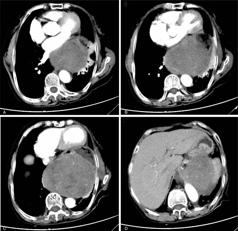
Figure 1.

Contrast-enhanced abdominal computer tomography (CT) showed a heterogeneous, 11.9 × 10.2 cm posterior mediastinal tumor in the left lower thorax, thrusting the distal esophagus, pylorus, cardiac forward, pressing the pulmonary leading to the atelectasis of part of the upper and inferior lobe of left lung, and adhering to the lower esophagus, pylorus, and the left adrenal gland with unclear boundaries.
