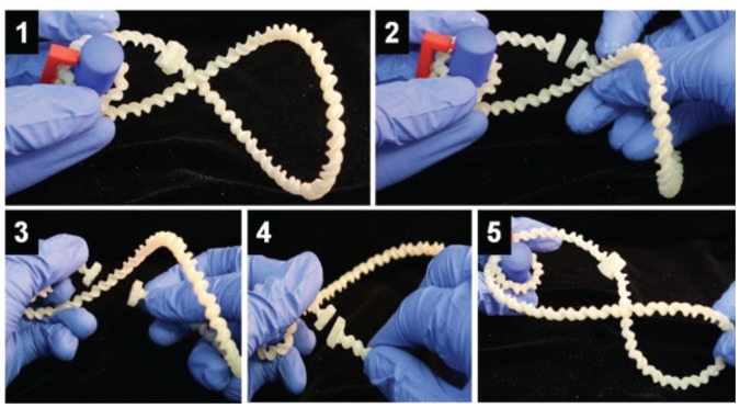
FIGURE 2.
Investigating DNA supercoiling. In step 1, students wrapped the DNA model (white) around a nucleosome model (blue) and characterized the resulting supercoil. In steps 2–4, students mimicked Topoisomerase II by cleaving the DNA and passing the intact strand of DNA through the cleaved site before re-adhering the ends. In step 5, students characterized the resulting supercoil and evaluated Topoisomerase II activity.

