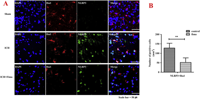
Fig. 6.
Fimasartan reduced activation of NLRP3 inflammasome largely in activated microglia within one day of ICH. Double labeling immunofluorescence staining images showed co-labeling for NLRP3 (A), and Iba-1 merged with DAPI around the hematoma in brain cryosections. Representative microphotographs show NLRP3 was activated in microglia cells labeled by Iba-1. Analysis of NLRP3 inflammasome immunoreactivity in the microglia expressed as a number of NLRP3 and Iba-1 positive cells (B). An increase in the proportion of the area positively stained with the Iba-1 antibody was used to measure microglia activation. Data are presented as mean ± SEM. Scale bar = 50 μm. n = 5 per group; ** p < .01.

