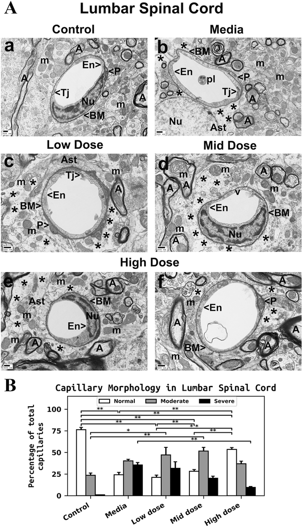
Figure 2. Characteristics of capillary ultrastructure in the lumbar spinal cord of G93A mice.
(A) Electron microscopy examination of microvasculature in the lumbar ventral horn. Control mouse demonstrated intact ultrastructural morphology of ventral horn capillaries (Aa). Capillary consisted of typical endothelium composed of a single layer of endothelial cells, pericytes, tight junction, surrounding basement membrane, and adjacent astrocyte end-feet. Mitochondria and myelinated axons were well preserved in neuropil. (Ab) Mediatreated mice demonstrated degenerated endothelial cells and astrocyte foot-processes. Perivascular protein-filled edema was determined in numerous capillaries. Swollen mitochondria were also observed in the neuropil and within axons. Myelin disruptions in surrounding axons were noted. ALS mice receiving the low or mid cell dose treatment demonstrated no improvement in capillary morphology. Swollen or vacuolated ECs in numerous capillaries were detected in low (Ac) or mid (Ad) cell dose treated mice. Significant perivascular edema was determined in these cell-treated mice. Mitochondria with disrupted cristae were seen in axon (top left, Ad) in addition to degenerated myelin in many axons. (Ae, Af) In ALS mice treated with high cell-dose, numerous capillaries demonstrated typical endothelium morphology. Normal ultrastructural morphologies of endothelial cells and pericytes, adjacent astrocyte end-feet, and preserved axonal myelin were observed. Only small areas of perivascular edema were seen. En – endothelial cell, Tj – tight junction, BM – basement membrane, P - pericyte, Ast – astrocyte, A – axon, m – mitochondrion, v – vacuole, pl – platelets, Nu – nuclei, * - perivascular edema. Scale bar in a-f is 500 nm. (B) Quantitative analysis of capillary morphology. Capillary profiles in the lumbar spinal cords were similar to those in the cervical spinal cords. Control mice showed a high percentage of capillaries with normal morphology and a low percentage of moderately impaired capillaries. Media-treated mice demonstrated a significant reduction of morphologically normal capillaries, an increase of capillaries with moderate impairment, and a high percentage of severely damaged microvessels. Steadily increasing percentages of capillaries with normal morphology were determined in cell-treated mice with increased cell dose. A significant increase of normal capillaries was demonstrated in mice receiving the high cell-dose compared to low or mid cell dose treated animals. The percentage of severely compromised capillaries was significantly reduced in mice treated with the high cell dose. The percentages of capillaries with moderate impairment were increased in low or mid cell-treated mice vs. media, whereas a minor percentage decrease was noted in mice receiving the high cell-dose. *p < 0.05, **p < 0.01.

