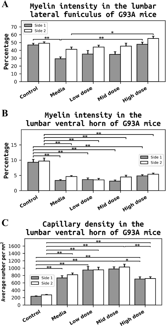
Figure 9. Quantitative analyses of myelin intensity and capillary density in the lumbar spinal cord of G93A mice.
(A) Media-treated ALS mice showed significant decrease of myelin in LF on side 1 of the lumbar spinal cord compared to control mice. There were no significant differences in myelin expressions in LF between the low or mid cell-dose treated mice vs. media. In contrast, mice treated with the high cell-dose demonstrated a significantly increased percentage of myelinated axons in LF on both sides of the lumbar spinal cord compared to media-treated mice. (B) Myelin intensity in VH was similar to LF. Significant decreases of myelin staining were determined in VH of media mice on both sides vs. controls. Although non-significant differences were found in VH myelin of the low or mid cell-dose treated mice vs. media, a trend towards a percentage increase of myelin expression was noted in mice after the high cell-dose treatment. (C) Quantitative analyses of VH of media-treated animals revealed significantly higher capillary densities on both sides of the lumbar spinal cord vs. controls. Continued elevations of capillary densities were also noted on both sides of VH in low and mid cell-dose treated animals compared to media ALS mice. Yet, a significant decrease of capillary density was determined in mice received the high cell-dose vs. mid cell-dose treated animals. *p < 0.05, **p < 0.01.

