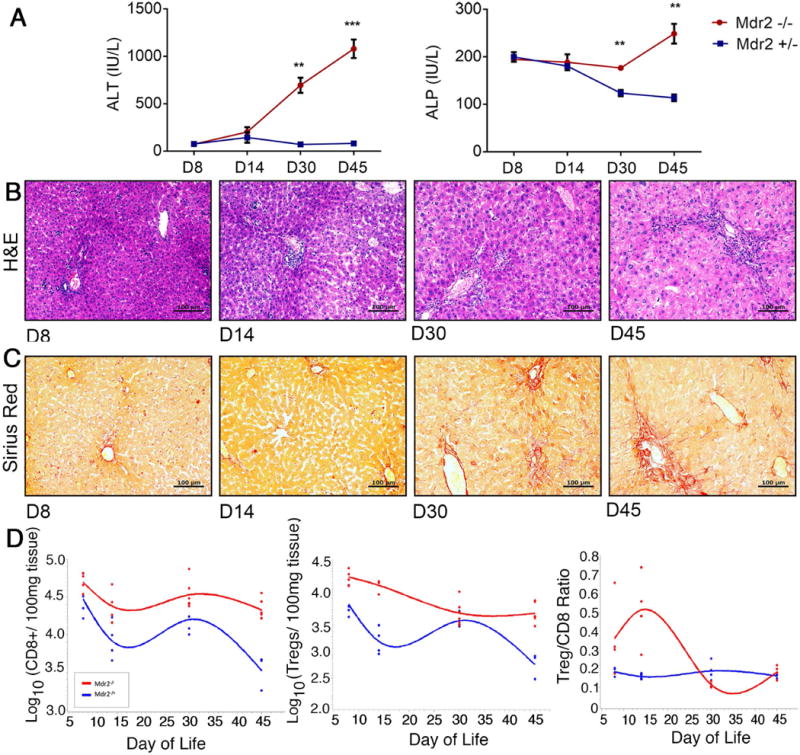
Fig. 1. Contraction of intrahepatic Treg lymphocytes coincides with hepatobiliary injury in Mdr2−/− mice.

(A) Plasma ALT and serum ALP concentrations were measured in FoxpP3-EGFP Mdr2−/− and age-matched control Foxp3-EGFP Mdr2+/- mice between 8 and 45 days of life using colorimetric assays (n=3-4/per group). Differences between groups were tested for statistical significance using an unpaired t test with **p<0.01 and ***p<0.005. In (B) and (C), representative photomicrographs of H&E and Sirius Red stained liver sections from Mdr2−/− mice demonstrate progression of periportal inflammation, bile duct proliferation and liver fibrosis. (D) Flow cytometry was performed on hepatic MNC from age-matched Mdr2−/− and +/- mice. Foxp3(GFP)+CD25+CD4+ Tregs and CD8+ T lymphocytes were enumerated within CD3+ size-gated lymphocytes (n=4-6/group), and total numbers of cells were log-transformed. Spline curves were generated to describe the kinetics of numeric Treg and CD8 lymphocyte responses in the liver during the first 45 days of life.
