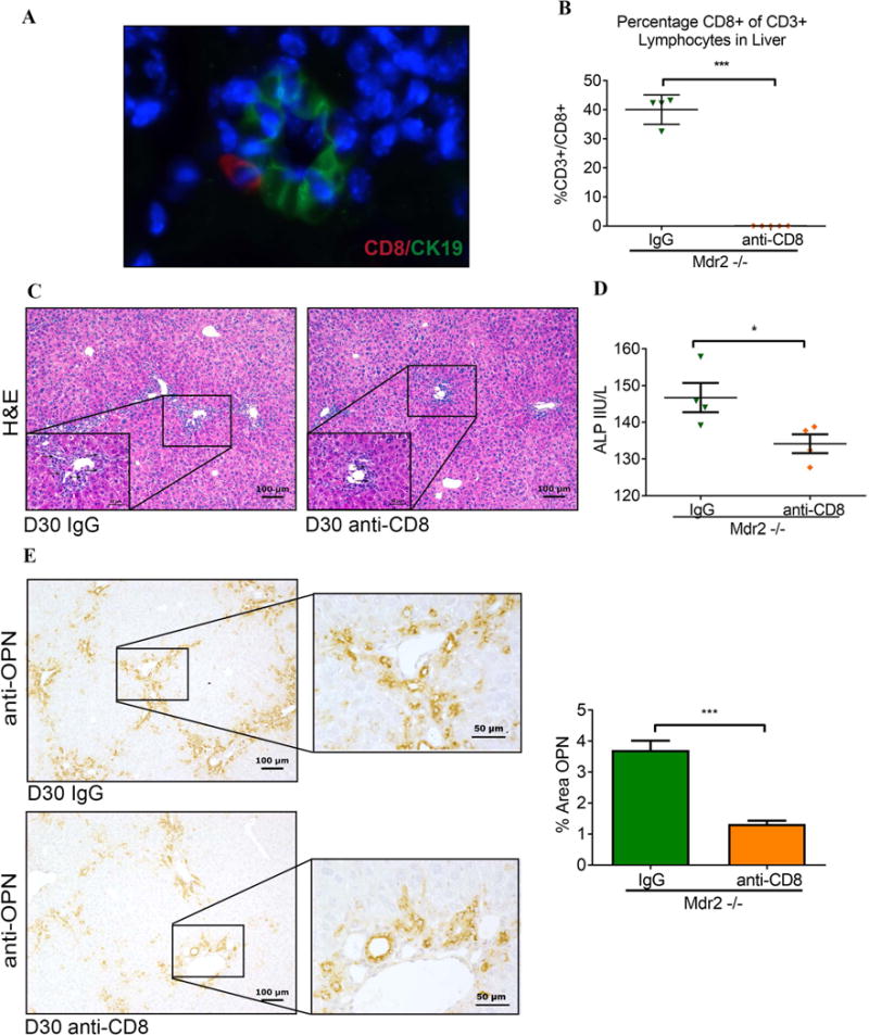
Fig. 2. Hepatic CD8+ T cells function as effector lymphocytes in “toxic bile” induced sclerosing cholangitis.

(A) Liver sections from 30-day-old Mdr2−/− mice were subjected to dual immunofluorescence with antibodies against CD8 and CK19. CD8+ cells (red) were detected infiltrating interlobular bile ducts identified by immunoreactivity against the cholangiocyte marker CK19+ (green). (B) Male Mdr2−/− mice were injected i.p. with anti-CD8 antibody or IgG isotype control twice weekly between day of life 14 and 30 and hepatic MNC from 30-day-old mice were subjected to flow cytometry. (C) Representative photomicrographs of H&E-stained liver sections demonstrate reduced bile duct proliferation in CD8-depleted Mdr2−/− mice (arrows denote bile duct profiles in high magnification inserts). (D) Serum ALP levels were measured on day 30 following anti-CD8 vs IgG treatment. (E) Liver sections from 30-day-old CD8-depleted and control Mdr2−/− mice underwent OPN immunohistochemistry as shown in representative photomicrographs. Immunoreactivity for OPN was quantified by automated image analysis (n=3 mice/group). Statistical analysis was performed by applying unpaired t test with *p<0.05 and ***p<0.005.
