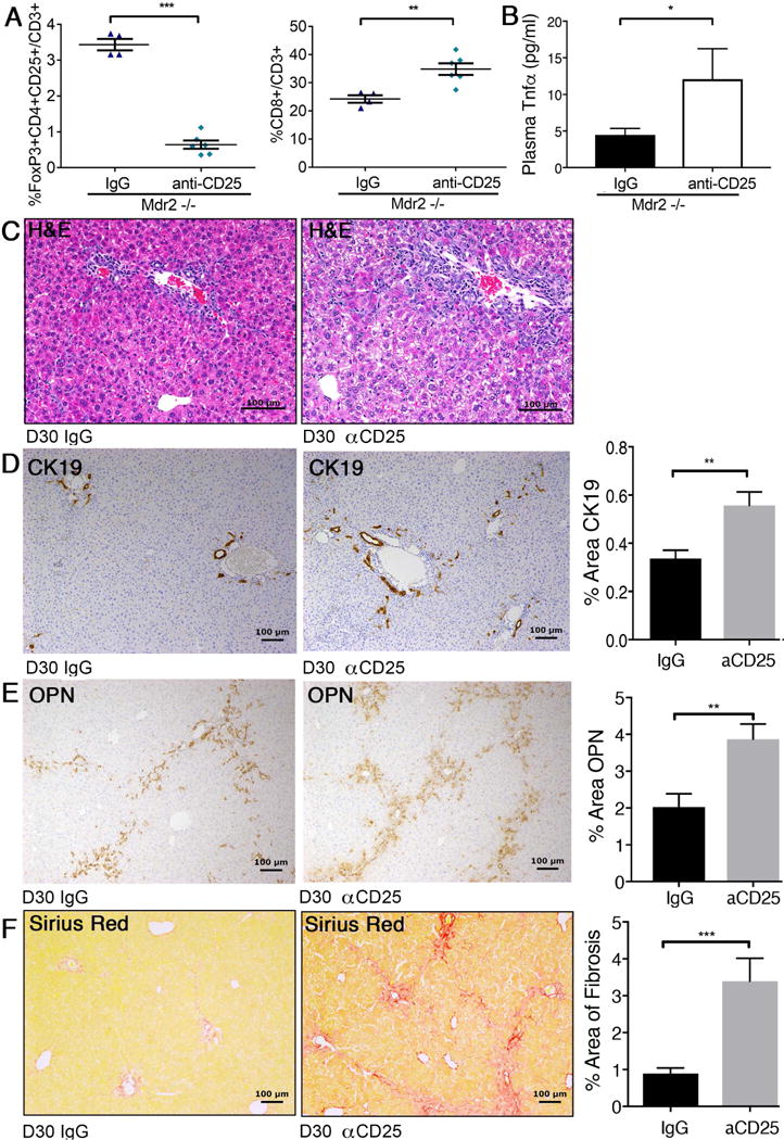
Fig. 3. Depletion of hepatic Tregs increases hepatic CD8+ lymphocytes, biliary injury, and fibrosis in Mdr2−/− mice.

Juvenile Mdr2−/− mice transgenic for FoxP3-EGFP received i.p. injections of CD25-depleting antibody or IgG isotype control between day of life 7 and 30. Hepatic MNC from 30-day-old mice of both groups were subjected to flow cytometry to enumerate populations of FoxP3+CD25+ Treg and CD8+ lymphocytes (A). Plasma concentrations for Tnfa were measured by Luminex (B). Liver histology was assessed by H&E-stained liver sections from CD25-depleted and control 30-day-old Mdr2−/− mice and revealed increased periportal inflammation in Treg-depleted mice as shown on representative photomicrographs (C). Liver sections from both groups of mice were subjected to IHC against CK19 (D) and osteopontin (E), and to Sirius Red staining to evaluate fibrosis (F). Immunoreactivity and collagen deposition were quantified by image analysis. Differences between groups were tested for statistical significance using unpaired t test with *p<0.05, **p<0.01 and ***p<0.005.
